Question
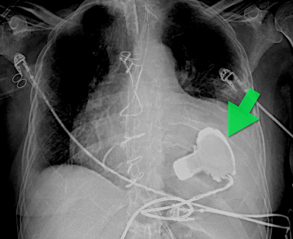
57 year old male presents with a cough. The CXR is shown below. What's the diagnosis?
Introduction
Fosphenytoin is a prodrug and is metabolized quickly to phenytoin after administration. The conversion of fosphenytoin to phenytoin involves the release of phosphate. In fact, each mmol of fosphenytoin releases 1 mmol of phosphate.
Clinical Question
Are patients at risk for hyperphosphatemia after fosphenytoin loading?
Data
There are only two cases of reported hyperphosphatemia.
Bottom Line
Despite the phosphate load from fosphenytoin administration, hyperphosphatemia is very rare and probably associated with renal insufficiency and dosing errors.
Acute ischemic stroke occurs in 3.3/100,000 children per year. Up to 30% of these are caused by varicella. This can be diagnosed if the patient has had varicella infection within the past 12 months, has a unilateral stenosis of a great vessel, and has a positive PCR or IgG from the CSF.
Treatment includes anticoagulation, acyclovir for at least 7 days and steroids for 3-5 days.
Outcome is normally good and spontaneous improvement can be seen.
Inflammation of other arteries, including other areas of the brain, can also be seen. Treatment options for this can include high dose glucocorticoids and possibly immunosuppresive agents.
Approval of Kcentra™ may open the door for studying treatment of the bleeding patient on newer oral anticoagulants.
General Information: Antibiotics are generally classified as time- and concentration-dependent.
Concentration-dependent antibiotics
-Fluoroquinolones (i.e. Levofloxacin)
-Aminoglycosides (i.e. Gentamicin)
-Azithromycin
Relevance to the EM Physician:
Concentration-dependent antibiotics should be given at the highest appropriate dose for the target tissues (i.e. Levofloxacin 750mg for pneumonia is preferable to 500mg). This is also the rationale for high dose, extended-interval dosing for Gentamicin (>5mg/kg initial dose).
University of Maryland Section of Global Emergency Health
Author: Andi Tenner, MD, MPH
Neuromuscular Blocking Agents in the Critically Ill
57 year old male presents with a cough. The CXR is shown below. What's the diagnosis?
You have a patient with a spinal cord syndrome and you order the MRI. Have you ever had that conversation with radiology where you have to "choose" what part of the spine you want imaged?
The entire spine needs to be imaged!
The reason: False localizing sensory levels.
For example: The patient has a thoracic sensory level that is caused by a cervical lesion.
A study of 324 episodes of malignant spinal cord compression (MSCC) found that clinical signs were very unreliable indicators of the level of compression. Only 53 patients (16%) had a sensory level that was within 3 vertebral levels of the level of compression demonstrated on MRI.
Further, pain (both midline back pain and radicular pain) was also a poor predictor of the level of compression.
Finally, of the 187 patients who had plain radiographs at the level of compression at referral, 60 showed vertebral collapse suggesting cord compression, but only 39 of these predicted the correct level of compression (i.e. only 20% of all radiographs correctly identified the level of compression).
The authors note that frequently only the lumbar spine was XR at the time of clinical presentation (usually at the referring hospital), presumably due to false localizing signs and a low awareness on the part of clinicians that most MSCC occurs in the thoracic spine (68% in this series).
An overweight 5 year old male presents with acute onset abdominal pain that localizes to the right lower quadrant. What are some causes of a limited or nondiagnostic ultrasound study in children?
Acute appendicitis is a time sensitive diagnosis. Ultrasound is frequently used as the initial diagnostic imaging in children. There are several reasons why the appendix may not be visualized, including retro-cecal location, normal appendix, perforation, and inflammation around the distal tip. An additional clinical predictor associated with poor or inconclusive ultrasound results in appendicitis is increased BMI (body mass index).
A study examining 263 pediatric patients found when BMI > 85th percentile and clinical probability of appendicitis was <50%, 58% of ultrasounds were nondiagnostic. Children with a BMI <85th percentile and clinical probability of appendicitis was <50%, had nondiagonstic scans 42% of the time. These trends were also mimicked in the patients with a higher clinical probability of appendicitis. In the child with a nondiagnostic ultrasound, options include observation and repeat ultrasound scan or CT scan, both of which have associated risks.
If you are working in a community hospital and have an acetaminophen overdose, one of the criteria to transfer the patient to a tertiary care center is presence of the King's College Criteria.
The below is taken from mdcalc.com - http://www.mdcalc.com/kings-college-criteria-for-acetaminophen-toxicity/
Each one is assigned points and can be prognostic for severe toxicity and need for transplant. The lactate and phosphorus are new ones and have modified the criteria. Phosphorus is utilized to create glycogen. If the liver is injured and trying to heal, your phosphorus will be low (good). If the liver is injured and unable to repair itself the phosphorus will be high (bad). This single test has an excellent prognostic ability.
| Lactate > 3.5 mg/dL (0.39 mmol/L) 4 hrs after early fluid resuscitation? | |
| pH < 7.30 or lactate > 3 mg/dL (0.33 mmol/L) after full fluid resuscitation at 12 hours | |
| INR > 6.5 (PTT > 100s) | |
| Creatinine > 3.4 mg/dL (300 µmol/L) | |
| Grade 3 or 4 Hepatic Encephalopathy? | |
| Phosphorus > 3.75 mg/dL (1.2 mmol/L) at 48 hours |
General Information:
The two main units used by medical laboratories are "conventional (used in the US) and SI (used by most other countries).
Pearls to know:
Relevance to the EM Physician:
These tips will help you convert labs to familiar values when reading medical literature, when working in another country, or when working with international colleagues.
University of Maryland Section of Global Emergency Health
Author: Andi Tenner, MD, MPH
Necrotizing fasciitis (NF) is a rapidly progressive bacterial infection of the fascia with secondary necrosis of the subcutaneous tissue. In severe cases, the underlying muscle (i.e., myositis) may be affected.
Risk factors for NF include immunosuppression (e.g., transplant patients), HIV/AIDS, diabetes, etc.
There are three categories of NF:
In the early stage of disease, diagnosis may be difficult; the physical exam sometimes does not reflect the severity of disease. Labs may be non-specific, but CT or MRI is important to diagnose and define the extent of the disease when planning surgical debridement.
Treatment should be aggressive and started as soon as the disease is suspected; this includes:
2013 AAP AOM Guidelines UPDATE
With recent events, a few notes about ricin seems appropriate:
CDC website: http://www.bt.cdc.gov/agent/ricin/
General Information:
A parasitic infection caused by the tissue-dwelling filarial nematode worm Wuchereria bancrofti; a wide range of mosquitoes transmit the infection. When the worm is mature, it inhabits lymph nodes and produces sheathed microfilarial larvae that circulate in the peripheral blood.
Clinical Presentation:
- Infection with the adult worms produces painless subcutaneous nodules that are usually less than 2 cm in diameter, typically over bony prominences.
- Symptoms depend on where the microfilariae migrate to, and vary accordingly. They include: pruritus, papular dermatitis, dermal atrophy and depigmentation or hyperreactive skin disease (Sowda), keratitis, iritis, chorioretinitis, optic atrophy and eventually blindness, orchitis, hydrocele, chyluria, elephantiasis, pulmonary eosinophilia, cough, wheezing, and splenomegaly.
Diagnosis:
- Peripheral blood smear taken between 11pm and 1am or after provocation using diethylcarbamazine (DEC).
- Filarial antigen test.
- Eosinophilia, and specific antiflarial IgG and IgE antibodies.
Treatment:
- DEC which must be obtained directly from the CDC.
- Alternatively Doxycycline. Both drugs are effective against both macro and micro-filaria.
Bottom Line:
One billion people globally are at risk for infection with filaria. 120 million already have the infection. Suspect the infection in patients that have been to Africa, Asia, especially India, Western pacific, Haiti, the Dominican Republic, Guyana and Brazil.
University of Maryland Section of Global Emergency Health
Author: Walid Hammad, MD
Massive Transfusion Pearls
35 year-old female presents with fever and hypotension. Bedside ultrasound is performed and is shown here. What's the diagnosis?
You have a patient with a spinal cord syndrome and you order the MRI. Have you ever had that conversation with radiology where you have to "choose" what part of the spine you want imaged?
The entire spine needs to be imaged!
The reason: False localizing sensory levels.
For example: The patient has a thoracic sensory level that is caused by a cervical lesion.
A study of 324 episodes of malignant spinal cord compression (MSCC) found that clinical signs were very unreliable indicators of the level of compression. Only 53 patients (16%) had a sensory level that was within 3 vertebral levels of the level of compression demonstrated on MRI.
Further, pain (both midline back pain and radicular pain) was also a poor predictor of the level of compression.
Finally, of the 187 patients who had plain radiographs at the level of compression at referral, 60 showed vertebral collapse suggesting cord compression, but only 39 of these predicted the correct level of compression (i.e. only 20% of all radiographs correctly identified the level of compression).
The authors note that frequently only the lumbar spine was XR at the time of clinical presentation (usually at the referring hospital), presumably due to false localizing signs and a low awareness on the part of clinicians that most MSCC occurs in the thoracic spine (68% in this series).
Adrenal insufficiency (AI) can be a life-threating condition and is classified as primary (failure of the adrenal gland) or secondary (failure of hypothalamic- pituitary axis).
Common causes of primary adrenal insufficiency include autoimmune destruction, infectious causes (TB and CMV), or interactions with drugs (e.g., anti-fungals, Etomidate, etc.). Secondary causes are usually due to abrupt withdrawal of steroids after chronic use, although sepsis and diseases of the hypothalamus or pituitary (e.g., CVA) may occur.
Signs and symptoms include fatigue, weakness, skin pigmentation, dizziness, abdominal pain, and orthostatic hypotension; it should be suspected with any of the following: hyponatremia, hyperkalemia, hypoglycemia, hypercalcemia, low free-cortisol level, and hemodynamic instability despite resuscitation.
Treatment:
• Correct underlying the disorder
• Resuscitation and hemodynamic support
• Correct hypoglycemia and electrolyte abnormalities
• Treat with hydrocortisone, cortisone, prednisone, or dexamethasone +/- fludrocortisone (Note: dexamethasone is attractive choice in the ED because it will not interfere with ACTH stimulation test)