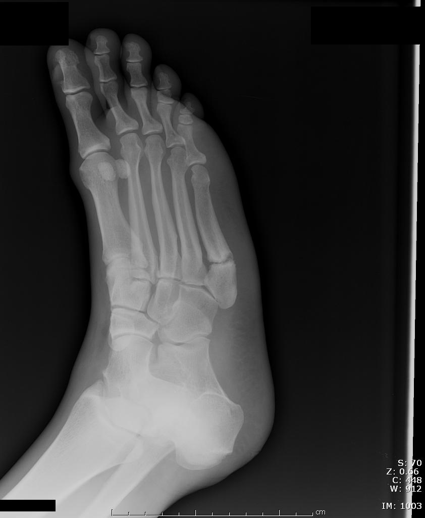
AC Joint Dislocations
The acromioclavicular (AC) Joint is commonly injured when a person falls onto their shoulder.
The AC Joint consists of three ligaments:
Injuries to this joint are classified as Type I – Type VI and involve sprain or tears of the AC or CC ligaments
Monteggia's Fracture
Jones fracture
Presented with persistant foot pain from
Jones fracture malunion.
Blast Injuries:
In honor of the 4th of July holiday, here is a quick pearl about blast injuries.
Metacarpal Fractures and Growth Plates:
The growth plates on metacarpals are on the distal end of the bone, except for the 1st metacarpal which is on the proximal end near the carpal bones.
Don't mistake this for a fracture line, however, make sure you get comparison views if they are tender over the area, as this can help you diagnosis a Salter Harris Type 1 fracture.
High Pressure Injection Injuries:
Shoulder Dislocations -- Treatment
Nursemaid Elbow:
It is typically taught that the way to reduce a nursemaid's elbow is to hold the elbow at 90 degrees, then firmly supinate and flex the elbow. Place your thumb over the radial head and apply pressure as you supinate.(Taken from Sean Fox's Pearl on 7/20/2007)
However, there is a growing body of evidence that is showing that hyperpronating the forearm actually has a higher success rate on first attempt, is easier to perform, and is associated with less pain then supinating the forearm. The overall reducation rates where similar for both methods.
The hyperpronation method consists of hyperpronating the forearm and then flexing the elbow. Since the child tends to already hold their arm in partial pronation, the hyperpronation technique tends to need less force and has been associated with less pain.
Elbow Dislocation
Quick clinical clues that the elbow is dislocated:
Trimallelor Fractures:
Bimallelor fracture involve both the medial mallelous of the tibia and the distal fibula. The third malleloi is the posterior tip of the articular surface of the tibia. Can result in instability in the posterior and lateral directions along with external rotation.
Some indications for Open Reduction Internal Fixation when the posterior mallelous is fractured are:
Knee Dislocations:
Are relatively rare injuries, but can result in loss of the limb if missed. Patients will sometimes say they dislocated their knee when they actually mean their patella, so a good history where they describe what their knee looked like, and what they were doing at the time will help differentiated the two.
Some signs that you are dealing with a spontanously reduced knee dislocation are:
The loss of limb is due to unrecognized injury to the popiteal artery which as be estimated to occur 7-45% of the time.
If you would like to see some videos of knee injuries in the making follow this link www.csmfoundation.org/Educational_Lower_Extremity.html
Distal Radius Fractures
Radial Head Fractures:
Radial head fractures are more common in adults, where radial neck fractures are more common in children. Remember to look for fat pads to help make the diagnosis if it is not obvious on plain films. On plain films, a line drawn down the middle of the radial head should always line up with the capitellum of the humerus. If this does not occur the radial head is dislocated and/or fracture.
Orthopaedics use the Mason classification to help guide treatment, and break down fractures into 3 different types.
Hamate Fractures:
Lunate Dislocation and perilunate dislocation are broken down into 4 stages that relates to the progressive disruption of the carpal ligaments due to hyperextension and ulnar deviation of the wrist:
For a good indepth review of lunate and perilunate injuries please read the article by Andy Perron with this attached link....
doi:10.1053/ajem.2001.21306
If you are interested in seeing some xray examples please visit LearningRadiology.com
A lot of what is taught about fracture patterns in abused children has been extrapolated from post-mortem studies which is a different population then what you will see in the Emergency Department. The study referenced did a metanalysis of all the literature in an attempt to determine what fractures suggest abuse and looked at all comers that had fractures. Some of the patterns they were able to extrapolate are:
The Galeazzi Fracture:
To see a photo of a Galeazzi fracture please visit the Learning Radiology Website by clicking on the following link:
http://www.learningradiology.com/caseofweek/caseoftheweekpix2/cow157lg.jpg
Most people are familiar with the Ottawa Ankle Rules, but there are also Ottawa Knee and Foot rules. The Ottawa rules help to limit the number of x-rays you may need in patients that present with ankle, foot or knee pain after an injury.
The Ottawa Ankle Rule
An ankle x-ray is only needed if there pain in the mallelolar area and any of the following:
The Ottawa Foot Rule
A foot x-ray is only needed if there is pain in the midfoot and any of the following:
The Ottawa Knee Rule
A knee x-ray is only needed for knee injury patients when they have any of the following:
Ankle sprains are typically treated with a short period of immbolization and then functional exercises are prescribed to rehabilitate the ankle. A study published in the Lancet this week might just change that. Lamb et al looked at 584 people with severe ankle sprains (unable to weight bear 3 days out from injury) that were randomized to be treated with a 10 day below knee cast, Aircast, Bledshoe Shoe or Tubular Compression dressing (similar to Ace Wrap). Those that were treated with the Cast and Aircast had quicker return to function and less disability at 3 months. There was no increased risk of DVTs in the cast group.
A commentary in the same issue points out that severe ankle sprains are associated with:
Based on this article I think it is prudent to treat all patients with severe Ankle Sprains with a prolonged period of forced immobilzation (Posterior Splint, Short Leg Cast or Aircast). I would also recommend the Aircast be used to prevent recurrent sprains especially if the patient is involved in sports that require jumping (Basketball, Volleyball) where the risk of reinjury is higher.