1-6 of 6 results by Cody Couperus-Mashewske


Title: Troubleshooting Ventilator Dyssynchrony

Category: Critical Care

Keywords: ventilation ineffective-trigger double-trigger (PubMed Search)

Posted: 6/24/2025 by Cody Couperus-Mashewske, MD

Patient-ventilator dyssynchrony is a sign of a disagreement between the patient's breathing and the ventilator's settings. Recognizing and fixing it is a critical skill to prevent lung injury and improve comfort. Ineffective triggering and double-trigger are two common types of dyssynchrony.

Ineffective Triggering

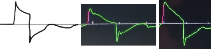
The patient tries to take a breath, but they are too weak to trigger the ventilator. This is the most common type of dyssynchrony. It causes increased work of breathing and discomfort.

Look for a small dip in the pressure waveform and a simultaneous scoop out of the expiratory flow waveform that is not followed by a delivered breath.

Troubleshooting options:

Double-Triggering ("Breath Stacking")

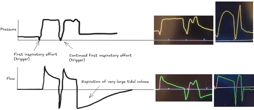
The patient's own breath outlasts the ventilator's set inspiratory time (Ti), causing one patient effort to trigger two stacked breaths. This results in delivery of large tidal volumes, risking lung injury (volutrauma).

Look for two consecutive breaths on the ventilator screen without a full exhalation in between.

Troubleshooting options:

Bottom Line

Dyssynchrony means the ventilator settings do not match the patient's needs. Watch the waveforms to diagnose the mismatch, then either adjust the ventilator or treat the underlying problem.

Show References


Title: HFNO vs. NIV in Respiratory Failure: The RENOVATE Trial

Category: Critical Care

Posted: 4/22/2025 by Cody Couperus-Mashewske, MD

This large RCT compared High-Flow Nasal Oxygen (HFNO) against Noninvasive Ventilation (NIV) via face mask in 5 types of Acute Respiratory Failure (ARF): non-immunocompromised hypoxemia, immunocompromised hypoxemia, COPD with acidosis, acute cardiogenic pulmonary edema (ACPE), and COVID-19.

Bottom Line:
RENOVATE suggests HFNO might be a reasonable, more comfortable initial choice for non-immunocompromised hypoxemic ARF or COVID-19 ARF. However, exercise caution using HFNO first-line for COPD exacerbations with acidosis or immunocompromised hypoxemic ARF due to conflicting analyses and potential harm signals. The signal for HFNO benefit in ACPE is intriguing but needs confirmation before changing practice. Close monitoring for failure and timely escalation are essential regardless of the initial noninvasive strategy.

Show References


Title: Troubleshooting Arterial Lines

Category: Critical Care

Posted: 2/18/2025 by Cody Couperus-Mashewske, MD

Arterial lines are essential tools in managing critically ill patients, but it is frustrating when they are not working as expected. It can be hard to tell when an unexpected waveform or pressure reflects the patient's physiology versus a problem with the line. Recognizing common issues and systematic troubleshooting will optimize your hemodynamic monitoring.

Types of arterial line problems

Troubleshooting Steps

  1. Correlate with Non-Invasive BP - MAPs should be within ~10 mmHg. Discrepancies suggest one of the numbers may be inaccurate. Make sure the cuff is the correct size!
  2. Verify Transducer Position - Level transducer at the 4th intercostal space, mid-axillary line. For each 10 cm off there is about 8 mmHg of pressure inaccuracy.
  3. Inspect Tubing and Pressure Bag
    • Ensure no kinks
    • Make sure the pressure bag is inflated to 300 mmHg
    • Flush vigorously to clear bubbles
  4. Check for Clots (radial lines):  Use ultrasound with Doppler to visualize flow and detect perica­theter clots. Reduce insonation angle (<60°) for optimal signal. “Positional” lines may have a clot around it, and the line only works well when it’s “hubbed” or the wrist is flexed.
  5. Consider exchanging the line over a micropuncture wire - it's more sterile and safest to place another line, but when access is tough/limited, it's not unreasonable to exchange a 4.45 cm 20g radial catheter for a 12 cm 20g catheter over a micropuncture wire with sterile technique.

By following these steps, you can systematically identify whether waveform or pressure abnormalities are due to technical issues or true patient physiology.

Show References


Title: Beyond the IVC: VExUS Exam Ultrasummary

Category: Critical Care

Keywords: VExUS, IVC, POCUS, Venous Congestion (PubMed Search)

Posted: 12/10/2024 by Cody Couperus-Mashewske, MD

The Venous Excess Ultrasound (VExUS) exam integrates IVC, portal, hepatic, and renal vein findings to assess venous congestion and guide management, such as diuresis, in critically ill patients.

Technique:

  1. IVC: Measure the IVC diameter. If <2 cm, significant congestion is unlikely, and further assessment is not well validated.
  2. Hepatic & Portal Veins: Use a curvilinear probe with color Doppler in the RUQ. The hepatic vein flows away from the probe (blue), and the portal vein, with thicker walls, flows toward the probe (red).
  3. Hepatic Vein Doppler: Apply pulse wave Doppler to the hepatic vein or a tributary. If the waveform is not clear, try a different vein.
  4. Portal Vein Doppler: After evaluating the hepatic vein, place PW Doppler on the portal vein.

Tips:

Interpretation:

Sometimes when other clinical information is contradictory, having the extra data point of the VExUS exam can be extremely useful to determine the best plan for a patient. Practice looking for the portal/hepatic veins and getting the waveforms on patients with a CLEAR clinical picture of venous congestion, then practice on more difficult cases.

Show References


Title: Using a Micropuncture Kit for Difficult Lines

Category: Critical Care

Keywords: vascular access, micropuncture kits, procedures (PubMed Search)

Posted: 10/15/2024 by Cody Couperus-Mashewske, MD

Getting reliable venous and arterial access is crucial when resuscitating critically ill patients. These lines can be difficult due to patient and situation specific variables. 

Micropuncture kits contain a 21-gauge echogenic needle, a stainless-steel hard shaft/soft-tip wire, and a 4 Fr or 5 Fr sheath and introducer. The micropuncture kit offers several advantages that can help overcome difficult situations:

To use a micropuncture kit, gain vessel access with the needle and wire, railroad the sheath and introducer into the vessel, remove the wire, then remove the introducer. Now you have a 4 Fr or 5 Fr sheath in the vessel. This is typically used to introduce a normal central line wire. 

For arterial lines, you can place them directly over the wire without dilation. Keep in mind that the 4 Fr sheath (1.3 mm OD) and 5 Fr sheath (1.7 mm OD) are larger than a typical arterial line catheter (18g = 1.27 mm OD). If you dilate then you will cause hematoma.

Find out where your department stores micropuncture kits and get familiar with their components. While it adds an extra step to the procedure, it could make the difference between securing the line or not.

Show References


Title: Euglycemic DKA Pitfalls and Pearls

Category: Critical Care

Keywords: Euglycemic DKA (PubMed Search)

Posted: 8/20/2024 by Cody Couperus-Mashewske, MD

Euglycemic DKA (eDKA) is a medical emergency requiring prompt attention. It is caused by an imbalance of insulin and glucagon leading to ketone accumulation (1-3). In addition to typical risk factors for DKA, those for eDKA include SGLT-2 inhibitor use and pregnancy, with 30% of DKA cases in pregnancy presenting euglycemic (4, 5).

eDKA presents with an anion gap metabolic acidosis, ketosis/ketonuria, & blood glucose less than 250 mg/dL.

Diagnosis requires ruling out other causes of anion gap metabolic acidosis, including toxic ingestions.

The cornerstone of eDKA management is ensuring enough dextrose to allow needed insulin administration to reverse ketone accumulation.

Pitfalls

Pearls

Show References