261-280 of 860 results with category "Critical Care"

Previous  |  1 |  ... |  9 |  10 |  11 |  12 |  13 |  14 |  15 |  16 |  17 |  18 |  19 |  ... |  43 |  Next

Title: Emergent TIPS as treatment for variceal bleeding

Category: Critical Care

Keywords: Cirrhosis, Varices, GI Bleeding, TIPS, Interventional Radiology (PubMed Search)

Posted: 12/17/2019 by Mark Sutherland, MD

There are few conditions that can be as dramatic or difficult to control as variceal GI bleeding in a cirrhotic patient.  It is important to be familiar with all options in these cases, from Blakemore/Minnesota tube placement to massive transfusion to when and which consultants to get involved.  In cases that are refractory or not amenable to endoscopic intervention, emergent interventional radiology consultation for Transjugular Intrahepatic Portosystemic Shunt (TIPS) may be a consideration.  In high risk cases, think about getting IR on the phone at the same time as you engage GI, in case endoscopic management fails.  Variceal bleed patients can decompensate rapidly, get your consultants involved early!


Generally accepted indications for emergent TIPS (both of the following should be true):

-GI bleeding not amenable or not controllable by endoscopy

-Cause is felt to be variceal. May also consider in portal hypertensive gastropathy


Contraindications:

-Right heart failure or pulmonary hypertension

-Severe liver failure (MELD > 22, T Bili > 3 or Child-Pugh C. In these cases TIPS may not confer a significant survival benefit)

-Hepatic encephalopathy (relative contradindication.  HE may be worsened by TIPS).

-Polycystic liver disease (makes TIPS technically challenging)

-Chronic portal vein thrombus (makes TIPS technically challenging. Acute PV thrombus is NOT considered a contraindication)


Bottom Line: In cases of variceal GI bleeding from portal hypertension, consider getting IR on the phone early to discuss emergent TIPS.

Show References


Title: DDAVP for intracranial hemorrhage

Category: Critical Care

Keywords: DDAVP, desmopressin, ICH, intracranial hemorrhage, stroke, CVA, hyponatremia (PubMed Search)

Posted: 12/10/2019 by Robert Brown, MD (Updated: 12/10/2019)

Pearl: consider desmopressin (DDAVP) for patients with an intracranial hemorrhage who are taking an antiplatelet. Caution, this is not for patients with an ischemic stroke with hemorrhagic conversion and it was not specifically evaluated for patients on anticoagulation or going to the OR with neurosurgery.

How strong is this evidence? International guidelines already give cautious approval for this practice, and now there is a retrospective review to support it. Though there were only 124 patients in the trial, the rate of hemorrhage expansion was much lower in the DDAVP group (10.9% vs 36.2%, P = .002) and there was no increased risk of hyponatremia (no events reported).

Show Additional Information

Show References


Title: Interventions Shown to Reduce Mortality in RCTs

Category: Critical Care

Posted: 12/3/2019 by Mike Winters, MBA, MD (Updated: 3/4/2026)

Interventions Shown to Reduce Mortality in RCTs

Show References


Title: Conservative oxygenation during mechanical ventilation

Category: Critical Care

Keywords: conservative oxygenation (PubMed Search)

Posted: 11/26/2019 by Quincy Tran, MD, PhD (Updated: 3/4/2026)

Settings

Study Results:

Discussion:

This study’s results differed from previous single center study (Girardis JAMA 2016) or meta analysis (Chu DK, Lancer 2018), which showed mortality benefit in patients with conservative oxygen (Girardis & Chu) and more ventilator-free days (Girardis).

Conclusion: Conservative oxygen did not significantly affect the ventilator free days of mechanically ventilated patients.

Show References


Title: PEA ... or is it?

Category: Critical Care

Keywords: OHCA, cardiac arrest, resuscitation, PEA, pesudo-PEA, pulseless electrical activity (PubMed Search)

Posted: 11/12/2019 by Kami Windsor, MD


When managing cardiac arrest, it is important to differentiate PEA, the presence of organized electrical activity without a pulse, from "pseudo-PEA,"where there is no pulse but there IS cardiac activity visualized on ultrasound. 


Why: 

How: 

What:


Bottom Line: Pseudo-PEA is different from PEA. Utilize POCUS during your cardiac arrests to identify it and to help diagnose reversible causes, and treat it as a profound shock state with the appropriate supportive measures, i.e. pressors or inotropy. 

Show References


Title:

Category: Critical Care

Keywords: Right Ventricle, RV Size (PubMed Search)

Posted: 11/5/2019 by Kim Boswell, MD

Rapid Assessment of the RV on Bedside Echo

There are several causes of acute RV dysfunction resulting in a patient presenting to the ER with unstable hemodynamics. Some of these include acute cor pulmonale, acute right sided myocardial infarction and acute submassive or massive pulmonary embolism. While bedside assessment of the LV function is often performed by the ED physician, simultaneous evaluation of the RV can provide crucial information that can help guide therapeutic decisions to prevent worsening of the patient’s clinical condition. A rough guideline to determine RV size and function is below using the apical 4 chamber view.

Normal RV size :            <2/3 the size of the LV

Mildly enlarged RV :       >2/3 the size of the LV, but not equal in size

Moderately enlarged RV:  RV size = LV size

Severely enlarged RV:      RV size > LV size

Patients who are found to have RV dilation should be given fluids in a judicious fashion as the RV is not tolerant of fluid overload. Early diagnosis of the cause of acute RV failure should be sought to guide definitive therapy, but early institution of inotropic support should be considered. Frequent reassessments of biventricular function during resuscitation should be performed.


Show References

Attachments


Title: Think Quick! Is Narrow vs Wide better than memorizing Hs and Ts?

Category: Critical Care

Keywords: Pseudo-PEA, Shock, Resuscitation (PubMed Search)

Posted: 10/29/2019 by Mark Sutherland, MD

Ever been in an acute rescucitation and found yourself unable to remember all of those famous ACLS Hs and Ts?  I know I have.  A few years ago Littman et al published an alternative approach to critically ill, hypotensive medical patients with non shockable rhythms.  Unfortunately, it seems like some of the enthusiasm for this approach has died down, but I still think it's something you're more likely to recall in a pinch than the Hs and Ts and is a better way of getting started with a hypotensive non-trauma patient.  And it's so simple you may actually remember it!


1) Look at the monitor.  Is the rhythm narrow or wide?  

2a) Narrow - more likely a mechanical problem (tamponade, tension PTX, autoPEEP, or PE). Give IVF and search for one of these causes (and correct it!).  Keep in mind that ultrasound can help you differentiate a lot of these.

2b) Wide - more likely a metabolic problem (hyperK, sodium channel blockade, etc*). Give empiric calcium, bicarb, and other therapies targeted for these problems (if desired) and get stat labs.


Take a minute and either go to this REBEL EM post:

https://rebelem.com/a-new-pulseless-electrical-activity-algorithm/

To review this, or look at the attached diagrams.  


*Dr. Mattu would want me to remind you that hyperkalemia IS a sodium channel poisoned state, so there's no need to think of these two separately

Show References

Attachments


Title: The Critically Ill Geriatric Patient with Sepsis

Category: Critical Care

Posted: 10/15/2019 by Mike Winters, MBA, MD (Updated: 3/4/2026)

The Critically Ill Geriatric Patient with Sepsis

Show References


Title: Targeted Temperature Management for Cardiac Arrest with Nonshockable Rhythm

Category: Critical Care

Keywords: cardiac arrest, hypothermia, nonshockable rhythm (PubMed Search)

Posted: 10/8/2019 by Quincy Tran, MD, PhD

Rationale: Data regarding temperature management in patients suffered from cardiac arrest with nonshockable rhythm was inconclusive.

Objective: whether moderate hypothermia at 33C, compared with normothermia at 37C would improve neurologic outcome in patients with coma after cardiac arrest with nonshockable rhythm.

Outcome: survival with favorable 90-day neurologic outcome (Cerebral Performance Category scale 1-2/5)

SummaryThere was higher percentage of patients achieving CPC 1-2 in the hypothermia group (10.2%) vs normothermia group (5.7%, Hazard Ratio 4.5, 95% CI 0.1-8.9, p=0.04)

This randomized multicenter trial involved 581 patients with cardiac arrest and nonshockable rhythm.  Hypothermia group included 284 patients vs. 297 in the normothermia group.  Median GCS at enrollment = 3.

Majority of patients was cooled with the use of a basic external cooling device: 37% for hypothermia and 50.8% for normothermia group.

There was higher percentage of patients achieving CPC 1-2 in the hypothermia group (10.2%) vs normothermia group (5.7%, Hazard Ratio 4.5, 95% CI 0.1-8.9, p=0.04)

Limitation:

A. The study used strict enrollment criteria:

  1. CPR initiation within 10 minutes;
  2. CPR to ROSC within 60 minutes;
  3. epinephrine or norepinephrine infusion at < 1 ug/kg/min;
  4. No Child-Pugh class C liver cirrhosis

B. normothermia group had higher proportion of patients with temperature at 38C.

C. Hypothermia group underwent temperature management of 56 hours vs. 48 hours for normothermia patients.

Take home points:

In a selected group of patients with cardiac arrest and nonshockable rhythm, moderate hypothermia at 33C may improve neurologic outcome.

Show References


Title: Critical Care Pearls for Blood Transfusions

Category: Critical Care

Posted: 10/1/2019 by Caleb Chan, MD (Updated: 3/4/2026)

Blood Transfusion Thresholds in Specific Populations

Sepsis - 7 g/dL

Acute Coronary Syndrome - no current specific recommendations pending further studies

Stable Cardiovascular Disease - 8 g/dL

Gastrointestinal Bleeds

Acute Neurologic Injury - Traumatic Brain Injury - 7 g/dL

Postpartum Hemorrhage - 1:1:1 ratio strategy

Show References


Title: Vaping Associated Pulmonary Illness (VAPI)

Category: Critical Care

Keywords: VAPI, acute respiratory failure, vaping, e-cigarettes, e-hookah, juul, pulmonary disease, acute lung diease, ARDS (PubMed Search)

Posted: 9/24/2019 by Kami Windsor, MD


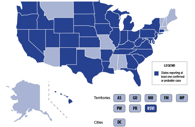
The U.S. is currently experiencing an epidemic of a severe lung disease termed Vaping-Associated Pulmonary Illness (VAPI), with over 500 cases and 7 deaths across 38 states and 1 U.S. territory since July 2019.

The clinical presentation of VAPI varies -- 

Diagnostics --

Treatment is supportive +/- steroids -- 


Bottom Line: Include vaping-associated pulmonary illness in your differential for patients presenting with acute lung disease.


Image result for vapi map vaping associated pulmonary illness


Show Additional Information

Show References


Title: Airway Management in the Critically Ill Pregnant Patient

Category: Critical Care

Keywords: Pregnant, difficult airway (PubMed Search)

Posted: 9/17/2019 by Kim Boswell, MD

Most non-OB physicians experience some fear or anxiety over taking care of the average pregnant patient. There are two patients to consider when caring for these women. Critical illness adds another layer of complexity to an already challenging patient population. Due to the normal physiologic changes that occur during pregnancy there are specific and important factors to be aware of when considering and preparing for intubation.


Show References


Title: VAD Troubleshooting

Category: Critical Care

Keywords: VAD, LVAD, Heart Failure (PubMed Search)

Posted: 9/10/2019 by Mark Sutherland, MD (Updated: 9/10/2019)

It's important to remember the differential for the patient with Ventricular Assist Device (VAD) difficulties, as these patients are likely to show up in your ED. 


1) Assess the patient as you usually would (signs of life, mental status, breathing, arrhythmias on monitor, etc). Listen for a hum over the chest.  Don't expect to feel a pulse.

2) Look at the VAD including controller, driveline, and power source for alarms, disconnections, signs of infection, and other obvious issues.

3) Look at the power (displayed flow), pulsatility index, and pump speed on the controller to help determine the cause of the issue (see attached chart).  Once you have a suspected etiology, typical management of these issues is usually similar to non-VAD patients (i.e. gentle IVF for hypovolemia, vasodilators if low flow is due to afterload/hypertension, defibrillation/CPR for arresting pts, etc).

Don't forget to call your VAD coordinator when able.  Consider a-line placement for precise evaluation of blood pressure (focus on MAP).


Bottom Line: Consider obstruction/thrombosis, bleeding, infection, hypovolemia, afterload/hypertension, arrhythmia, worsening LV function, and suction events when troubleshooting VADs.  The power, pulsatility index, and pump speed help differentiate these conditions.

Show References

Attachments


Title: Atrial Fibrillation in Critically Ill Patients

Category: Critical Care

Keywords: Atrial Fibrillation, sepsis, critical care, cardioversion, beta blockers, calcium channel blockers, rate control, rhythm control (PubMed Search)

Posted: 9/3/2019 by Robert Brown, MD (Updated: 3/4/2026)

One third of your critically ill patients will have atrial fibrillation. 

More than one third of those patients will develop immediate hypotension because of it.

More than one in ten will develop ischemia or heart failure because of it.

This is what you should know for your next shift:

#1 Don't wait to use electricity. If your patient is hypotensive or ischemic because of atrial fibrillation, you do not need to wait for anticoagulation before you cardiovert.

#2 Electricity buys you time to load meds. Fewer than half of patients you cardiovert will be in sinus rhythm an hour later and fewer than a quarter at the end of a day.

#3 There is no perfect rate control agent. Beta blockers have a lower mortality in A-fib from sepsis. Esmolol has the benefit of being short-acting if you cause hypotension. Diltiazem has better sustained control than amiodarone or digoxin. 

#4 There is no perfect rhythm control agent. Magnesium is first-line in guidelines. Amiodarone can be used even when there is coronary artery or structural heart disease.

#5 Anticoagulation is controversial. In sepsis, anticoagulation does not reduce the rate of in-hospital stroke, but does increase the risk of bleeding. Use with caution if cardioversion isn't planned.


Show Additional Information

Show References


Title: Critical Care Pearls for AIS

Category: Critical Care

Posted: 8/27/2019 by Mike Winters, MBA, MD

Critical Care Management of AIS

Show References


Title: Torsades de pointes and QT prolongation Associated with Antibiotics

Category: Critical Care

Keywords: Torsades de pointes, QT prolongation, antibiotics (PubMed Search)

Posted: 8/20/2019 by Quincy Tran, MD, PhD (Updated: 3/4/2026)

A new study confirmed the previously-known antibiotics to be associated with Torsades de pointes and QT prolongation (Macrolides, Linezolid, Imipenem and Fluoroquinolones). However, this study found new association between amikacin and Torsades de pointes/QT prolongation.

Methods

The authors queried the United States FDA Adverse Event Reporting System (FAERS) from 01/01/2015 to 12/31/2017 for reports of Torsade de points/QT prolongation (TdP/QT).

Reporting Odd Ratio (ROR) was calculated as the ratio of the odds of reporting TdP/QTP versus all other ADRs for a given drug, compared with these reporting odds for all other drugs present in FAERS

Results

FAERS contained 2,042,801 reports from January 1, 2015 to December 31, 2017. There were 3,960 TdP/QTP reports from the study period (0.19%).


Macrolides               ROR 14 (95% CI 11.8-17.38)

Linezolid                  ROR 12 (95% CI 8.5-18)

Amikacin                 ROR 11.8 (5.57-24.97)

Imipenem-cilastatin ROR 6.6 (3.13-13.9)

Fluoroquinolones   ROR 5.68 (95% CI 4.78-6.76)


Limitations:

These adverse events are voluntary reports

There might be other confounded by concomitant drugs such as ondansetron, azole anti-fungals, antipsychotics.

Show References


Title:

Category: Critical Care

Keywords: amikacin, Torsades de pointes, QT prolongation (PubMed Search)

Posted: 8/20/2019 by Quincy Tran, MD, PhD

Torsades de pointes and QT prolongation Associated with Antibiotics


Methods

The authors queried the United States FDA Adverse Event Reporting System (FAERS) from 01/01/2015 to 12/31/2017 for reports of Torsade de points/QT prolongation (TdP/QT).

Reporting Odd Ratio (ROR) was calculated as the ratio of the odds of reporting TdP/QTP versus all other ADRs for a given drug, compared with these reporting odds for all other drugs present in FAERS

Results

FAERS contained 2,042,801 reports from January 1, 2015 to December 31, 2017. There were 3,960 TdP/QTP reports from the study period (0.19%).


Macrolides               ROR 14 (95% CI 11.8-17.38)

Linezolid                  ROR 12 (95% CI 8.5-18)

Amikacin                 ROR 11.8 (5.57-24.97)

Imipenem-cilastatin ROR 6.6 (3.13-13.9)

Fluoroquinolones   ROR 5.68 (95% CI 4.78-6.76)


Limitations:

These adverse events are voluntary reports

There might be other confounded by concomitant drugs such as ondansetron, azole anti-fungals, antipsychotics.


Bottom Line:

This study confimed the previously-known antibiotics to be associated with Torsades de pointes and QT prolongation (Macrolides, Linezolid, Imipenem and Fluoroquinolones). However, this study  found new association between amikacin and Torsades de pointes/QT prolongation.

Show References


Title: Tips for Kidney Transplant Patients

Category: Critical Care

Posted: 8/14/2019 by Caleb Chan, MD

The Kidney Transplant Patient in Your ED

Show References


Title: Vent Management in Severe Obstructive Lung Disease

Category: Critical Care

Keywords: mechanical ventilation, respiratory failure, obstructive lung disease, asthma exacerbation, COPD (PubMed Search)

Posted: 8/6/2019 by Kami Windsor, MD


Managing the intubated patient with exacerbation of severe obstructive lung disease, especially asthma, can be very challenging as it carries higher risks of barotrauma due to higher pulmonary pressures and circulatory collapse due to auto-PEEP and decreased venous return. When measures such as medical therapy and noninvasive positive-pressure ventilation fail to prevent intubation, here are some tips to help:


1. Utilize a volume control ventilation mode to ensure a set tidal volume delivery / minute ventilation, as pressure-targeted modes will be more difficult due to the high pulmonary pressures in acute obstructive lung disease.

2. Set a low RR in order to allow for full exhalation, avoiding air-trapping / breath-stacking and circulatory collapse due to decreased venous return. This may require deep sedation and potentially paralysis.

3. Increase your inspiratory flow by shortening your inspiratory time (thereby increasing your time for exhalation.

4. Monitor for auto-PEEP:

5. Peak inspiratory pressures will be high -- what is more important is the plateau pressure, measured by performing an inspiratory hold at the end of inspiration. Provided your plateau pressure remains <30, you don't need to worry as much about the peak pressure alarms.

6. If your patient acutely decompensates in terms of hemodynamics and oxygenation -- first attempt to decompress their likely auto-PEEPed lungs by popping them off the ventilator and manually press on their chest to assist with exhalation of stacked breaths allowing venous return to the heart.


Show References


Title: Extubation Criteria

Category: Critical Care

Keywords: Mechanical Ventilation, Intubation, Extubation, RSBI (PubMed Search)

Posted: 7/30/2019 by Mark Sutherland, MD (Updated: 7/30/2019)

With increasing critical care boarding and the opioid crisis leading to more intubations for overdose, extubation - which was once a very rare event in the ED - is taking place downstairs more often.  Prolonged mechanical ventilation is associated with a ton of complications, so it's important for the ED physician to be comfortable assessing extubation readiness.  There is no single accepted set of criteria, but most commonly used are some variant of the following:

If the above criteria are met, two additional tests are frequently considered:

And don't forget to consider extubating high risk patients directly to BiPAP or HFNC!


Bottom Line: For conditions requiring intubation where significant clinical improvement may be expected while in the ED (e.g. overdose, flash pulmonary edema, etc), be vigilant about, and have a system for, assessing readiness for extubation.

Show References


Previous  |  1 |  ... |  9 |  10 |  11 |  12 |  13 |  14 |  15 |  16 |  17 |  18 |  19 |  ... |  43 |  Next