Injury was originally described as an occupational hazard in Scottish gamekeepers (from breaking the necks of rabbits against the ground). Today, skiing is now the most common cause and injury is now the second most common orthopedic injury in skiers (MCL injury #1).
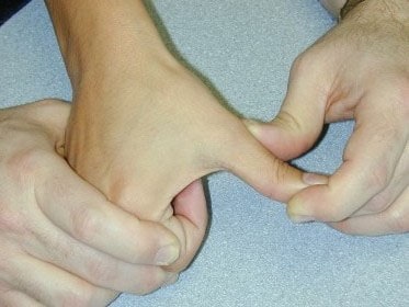
Injury to the ulnar collateral ligament (UCL) results from a sudden forced abduction (radial deviation) stress at the MCP joint of the thumb, commonly due to a fall against a ski pole or the ground.

http://blog.fitter1.com/wp-content/uploads/2010/04/b_14_1_2a.jpg
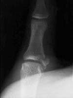
The most frequent site of rupture is the insertion into the proximal phalanx. The UCL may even avulse a small portion of the proximal phalanx at its insertion site.

http://img.medscape.com/pi/emed/ckb/sports_medicine/84611-97564-98460-1652013.jpg
Consider imaging before stress testing (to avoid further displacing a fracture)

http://img.medscape.com/pi/emed/ckb/sports_medicine/84611-97564-98460-1652060.jpg
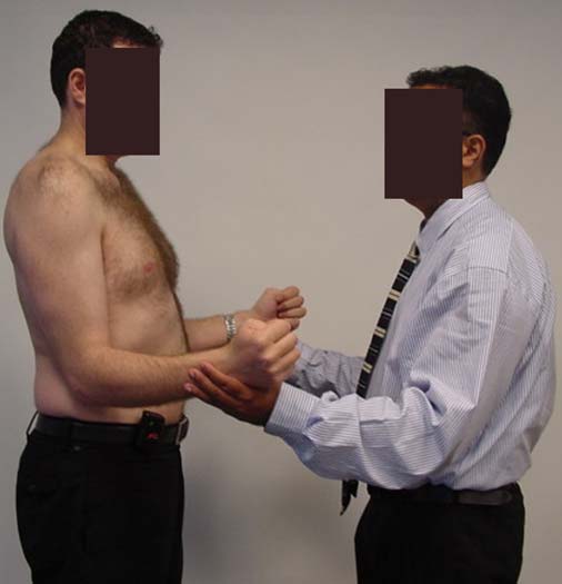
Stabilize in a thumb spica splint and refer to hand surgery.
Calling this entity a “simple sprain” may result in chronic disability (chronic pain, instability, loss of pinch strength)
Supraspinatus: “Empty can” test. Have the patient abduct the shoulders to 90 degrees in forward flexion with the thumbs pointing downward. The patient attempts to lift the arms against the examiner’s resistance.
http://bjsportmed.com/content/42/8/628/F2.large.jpg
Infraspinatus and teres minor: These muscles are responsible for external rotation of the shoulder. Have the patient flex both elbows to 90 degrees while the examiner provides resistance against external rotation.
http://www.physio-pedia.com/images/4/4b/Infraspinatus_test.jpg
Subscapularis: “Lift-off” test. The patient rests the dorsum of the hand on the lower back (palm out) and then attempts to move the arm and hand off the back. Patients with tears may be unable to complete test due to pain.
http://www.aafp.org/afp/2008/0215/afp20080215p453-f4.jpg
Radiologic evaluation of the elbow (Part 2)
Helpful clues in the evaluation of elbow trauma:
Helpful clues in the evaluation of elbow trauma
Fat pads: The fat pad sign can be seen with any joint effusion (infection, inflammation) but in the setting of trauma, effusions are indicative of fractures about the elbow (even if no fracture line can be identified).
There are two fat pads within the elbow. Normally, on a true lateral radiograph only the anterior fat pad is seen as a small triangular radiolucent shadow anterior to the distal humeral diaphysis. The posterior fat pad is ordinarily not visualized on a lateral radiograph because it is tucked away within the olecranon fossa.
Normal lateral view: http://nypemergency.org/images/ElbowNormal.jpg
With fractures, the joint becomes distended with blood. The anterior fat pad becomes displaced superiorly and outward from the humerus giving the so called "sail sign." Similarly, the posterior fat pad gets displaced out of the olecranon fossa and becomes visible on the lateral radiograph.
Anterior (sail) and posterior fat signs: http://nypemergency.org/images/Elbowsfatpadarrow.jpg
| History and Physical Examination Red Flags | |
| Historical Red Flags | Physcial Red Flags |
| Age under 18 or over 50 Pain lasting more than 6 weeks History of cancer Fever and chills Night sweats, unexplained weight loss Recent bacterial infection Unremitting pain despite rest and analgesics Night pain Intravenous drug users, immunocompromised Major trauma Minor trauma in the elder | Fever Writhing in pain Bowel or bladder incontinence Saddle anesthesia Decreased or absent anal sphincter tone erianal or perineal sensory loss Severe or progressive neurologic defect Major motor weakness |
http://www.gentili.net/signs/images/400/spinescottyparsdefectdrawing.JPG
The Scotty dog’s head (superior articular facet), nose (transverse process), eye (pedicle), neck (pars interarticularis), and body (lamina) should be easily identified on the oblique radiograph.